Hepatology, March 1999, p. 621-623, Vol. 29, No. 3
HEPATOLOGY Concise Review
Homeostatic Control of Angiogenesis: A Newly Identified Function of the Liver?
Bruno Clément,
From INSERM U-456, Detoxication and Tissue Repair Unit, University of Rennes I, Rennes, France.
Plasma proteins are produced and secreted at high levels by hepatocytes. The most abundant is albumin. The remainder are a heterogeneous group of glycoproteins, several of which have important homeostatic functions. This review will focus on two plasma proteins whose physiological effects expand the already extensive functionalrepertoire of liver-secreted proteins: plasminogen, which modulates breakdown of extracellular matrix (ECM) through its activation to plasmin; and collagen XVIII, which is a basement-membrane protein. Recent studies indicate that plasminogen is the precursor of angiostatin and collagen XVIII is the precursor of endostatin. Both angiostatin and endostatin are polypeptides that inhibit endothelial cell proliferation, angiogenesis, and tumor growth in experimental models of cancer. Hepatocytes appear to be the main source of these proteins, normally synthesizing and secreting them into plasma, and thus may influence extrahepatic endothelial growth.
ANGIOGENESIS
Many physiological and pathological processes require the formation of new blood vessels, a process known as angiogenesis. This is tightly regulated: blood vessels supply growing tissue with nutrients, growth factors, hormones, and oxygen but only to match the needs of development or repair of injured tissues. Tumors also depend on angiogenesis.1 Neovascularization isrequired for tumor growth and for migration of metastatic cells.When angiogenesis is reduced, growth and metastasis decreasesignificantly.
Angiogenesis occurs through endothelial sprouting from existing small vessels in response to a signal(s) from the tissue that requires a blood supply. Endothelial cells must first breach the basement membrane that surrounds existing vessels; they do this by elaborating proteinases, in cooperation with adjacent myofibroblasts, that break down the periendothelial basement membrane. Proliferationensues regulated by various stimulatory and inhibitory factors.2 Prominent among the proangiogenic factors are vascular endothelial growth factor, also known as vascular permeability factor, and fibroblast growth factors. These are present in most highly vascularizedtumors.3,4 Inhibition of endothelial cell growth and migration depends on a variety of proteins, including thrombospondin-1,SPARC/BM-40/osteonectin, platelet factor 4, prolactin, fibronectin fragments, angiostatin, and endostatin.5,6
PLASMINOGEN-ANGIOSTATIN
Plasminogen is secreted by hepatocytes and present in plasma and interstitial fluids at a concentration of 1 to 2 µmol/L. It is cleaved into plasmin through urokinase- and tissue-typeplasminogen activators.7 Plasmin is a serine proteinase that is broadly active: the classical substrates include fibrin, a major component of clots in leaky or damaged blood vessels; in addition, fibronectin, whose plasma form is produced by hepatocytes,8 laminin, and other ECM proteins are cleaved by plasmin during the remodeling of ECM that occurs in angiogenesis. Of interest, fibronectin-derived peptides strikingly inhibit endothelial cell proliferation in vitro.9
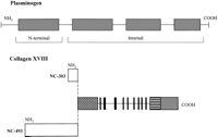
The first four domains of plasminogen, which are kringles, yield fragments that inhibit angiogenesis, one of which corresponds to the recently identified Mr=38-kd peptide, angiostatin (Fig.1).10-12 Different members of the ECM metalloproteinase family, including MMP3 (stromelysin), MMP7 (matrilysin), MMP9 (gelatinase B), and metalloelastase generate these fragments.10-12 Angiostatin is a potent inhibitor of angiogenesis in experimental models oftumors.13 It blocks metastatic murine Lewis lung carcinoma. Interestingly, the tumor cells appear to not express angiostatin; rather, the inhibitor derives from circulating plasminogen.14 Angiostatin also suppresses nontumor angiogenesis, in both the chick chorioallantoic membrane assay and in the mouse cornealassay,13 and in vitro it inhibits endothelial cell proliferation. The latter effect appears to be cell specific.13
| Fig. 1. Schematic representation of plasminogen and collagen XVIII and their cryptic fragments with angiogenesis inhibitory activities. Plasminogen: The N-terminal and internal fragments of angiostatin (gray boxes) include the first four of the five disulfide-linked structures of plasminogen, known as kringle structures. Collagen XVIII: The two N-terminal end variants of human collagen XVIII have noncollagenous domains fo 493 (NC-493) and 303 (NC-303) amino acids, respectively. Human hepatocytes express the NC-493 variant at high levels. Both variants share the C-terminal residues of the N-terminal NC domain (stippled box), the collagenous domains (full line), the noncollagenous interruptions (black boxes), and the C-terminal noncollagenous domain of the molecule (horizontal lines) containing endostatin (gray box). |
COLLAGEN XVIII-ENDOSTATIN
Collagen XVIII is a recently identified nonfibrillar collagen, forming together with collagen XV the MULTIPLEXIN (multiple triple-helix domains and interruptions) subgroup within the collagensuperfamily.15 The characteristics of these proteins are a large globular N-terminus, a highly interrupted triple-helical region, and a globular C-terminus with four conserved cysteines. The liver is an important site of collagen XVIII production.16 Variant forms appear to exist that differ with respect to the length of the noncollagenous (NC) N-terminal region, containing 303 (NC-303) or 493 (NC-493) residues17,18 (Fig. 1). One is associated with basement membrane,16,19 and one circulates in plasma.20
Endostatin is an approximately Mr=20-kd fragment of the C-terminal domain (Fig. 1). It has been found to inhibit angiogenesisand tumor growth in mice as well as the proliferation of endothelialcells in vitro.21 In four types of subcutaneous tumors, endostatin reduced tumor volume by 99%. Repeated cycles of administration caused total regression without evidence of resistance.22 Proteolyticrelease of endostatin and/or whole C-terminal noncollagenous domainof collagen XVIII can occur through several pathways yieldingan array of polypeptides ranging from Mr=38 kd to Mr=22 kd. Interestingly, the Mr=38-kd domain exhibits a higher affinity than does the Mr=22-kd protein for sulfatides and basement membrane proteins (laminin-1 and perlecan).23,24 These and other ECM-binding activities were predicted by the crystal structure of recombinant mouse endostatin23 and determined by binding assays.24 Tissue extracts contain high amounts of endostatin polypeptide with a molecular weight similar to the whole C-terminal domain; mouse liver has 2 µg/100 mg tissue.24 Serum samples from healthy humans contain variant forms similar to Mr=22-kd endostatin.20,24Immunological assays have shown that the concentration of endostatinin plasma is 120 to 300 ng/mL.24 The difference in concentration between tissue and serum suggests the existence in tissue of a pool of endostatin, possibly in association with ECM proteins. Proteolysis of this material may yield the more soluble forms found in serum.24
BIOLOGICAL ACTIONS
The way in which angiostatin and endostatin inhibit endothelial cell growth is unknown, but may resemble that of fibronectin fragments, which are postulated to act by binding to heparin and heparan sulfate.9 Consistent with this, the crystal structure of endostatin suggests the presence of a heparin-binding domain.23 Thus, both fibronectin- and collagen XVIII-derived polypeptides may block cell growth by competing with basic fibroblast growth factor for cellular heparan sulfate receptors. In addition, endostatin or another fragment(s) of collagen XVIII could compete for laminin, perlecan and fibulins, which are known to associate with fibronectin,24 disrupting endothelial cell interactions with basement membrane and promoting apoptosis. Whether ECM binding affects the biological activity of these polypeptides is unknown, i.e., whether they are active when bound or only after their release from ECM. In addition, although several forms of endostatin have similar properties, it is as yet unclear whether they exert similar biological effects.
Endostatin and angiostatin were identified originally in murine tumors. However, precursor forms are present in stromal cells, and circulating forms can be detected in high concentration.11,13,20,24It is not known whether the plasma forms are biologically active;activation may require local mechanisms, possibly cell-associatedproteinases. Production of plasminogen-angiostatin and collagenXVIII-endostatin by hepatocytes varies in liver disease,5,16,25,26potentially with consequences for extrahepatic endothelial cellproliferation and angiogenesis in development, tissue repair,and tumor growth. Interestingly, hepatic production of NC C-terminalpeptides of collagen XVIII containing the endostatin region appearsto be very low in highly angiogenic hepatocellular carcinomas(Musso et al., manuscript in preparation).25
It is currently speculated that mouse tumors treated with endostatin or angiostatin undergo apoptosis and necrosis because of hypoxia and nutrient deprivation; they appear to become completely dormant after several treatment cycles. This does not represent induction of an altered immune state, because a fresh inoculum of cancer cells at a different site still results in tumor formation. Rather, tumor dormancy likely reflects a high concentration of ECM-bound endostatin at the site of injection in the primary tumor.27 The local concentration may increase as the tumor shrinks 1/10 to 1/50 its original size and ECM condenses.
In conclusion, angiostatin and endostatin arise from proteins produced in the liver. They are examples of biological response modifiers with functions that appear to be completely different from those of the parent molecule. Their regulation also may differ from that of the parent. Their actual release requires proteolyticprocessing of plasminogen or collagen XVIII, and thus their fineregulation may depend on the expression of proteinase(s), whichin turn may be subject to unique regulatory factors. However,given that hepatocytes are the source of much of the precursorpool, regulation of angiogenesis may be regarded as a new liverfunction with important consequences for tissue repair andcancer.
Acknowledgment
The authors thank Pr. M. Bourel and A. Guillouzo, who initially stimulated our interest in hepatology, for critical review of the manuscript, and Pr. T. Pihlajaniemi for helpful discussions.This review is dedicated to the memory of André Clément.
Abbreviations
Abbreviations: ECM, extracellular matrix; NC, noncollagenous.
FOOTNOTES
Received October 23, 1998; accepted December 30, 1998.
Address reprint requests to: Bruno Clément, Ph.D., INSERM U-456, Facultés de Médecine et de Pharmacie, Rennes I University, 2 avenue Léon Bernard, 35043 Rennes, France. E-mail: bruno.clement@rennes.inserm.fr; fax: (33) 2 99 33 62 42.
REFERENCES
Copyright © 1999 by the American Association for the Study of Liver Diseases


