Management of Ectopic Varices
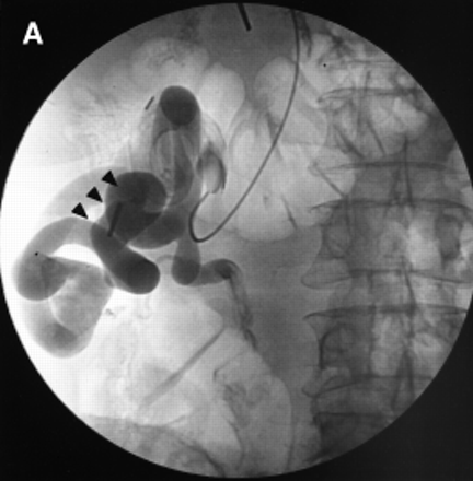
Fig. 2. (A) Angiographic view of the colonic varices (arrowheads) shown in Fig. 1. The transhepatic catheter is in the feeding mesenteric vein branch. (B) A late image from the same series shows the large draining systemic vein. (C) Angiography shows stasis in the feeding vessels (arrow) and disappearance of the varices.
 
|


