Hepatology, November 1998, p. 1173-1176, Vol. 28, No. 5
HEPATOLOGY Concise Review
A Tale of Two Strands: Reverse-Transcriptase Polymerase Chain Reaction Detection of Hepatitis C Virus Replication
David V. Sangar and A. R. Carroll
From the Department of Microbiology and Immunology, University of Texas Medical Branch at Galveston, Galveston, TX, and Glaxo Wellcome Research and Development, Medicines Research Center, Hertfordshire, UK.
“It was the best of times, it was the worst of times . . . . .” This phrase from Charles Dickens aptly describes the impact of the polymerase chain reaction (PCR) on Hepatitis C research. Clearly, the combination of reverse-transcriptase polymerase chain reaction (RT-PCR) and PCR amplification of viral complementary DNA (cDNA) has had a pivotal role in the identification of the virus responsible for the majority of non-A non-B hepatitis cases. However, although it has allowed the molecular characterization of this virus, the unique sensitivity of RT-PCR has also created a number of problems. For example, a cell culture system for the propagation of Hepatitis C virus (HCV) would be of great benefit. However, formal proof of replication in such systems has not been easy to obtain. The use of “strand-specific” RT-PCR to detect the replicative intermediates (minus-strand RNA) of HCV has been taken as evidence for viral replication. However, recent developments have shown that strand-specific RT-PCR is fraught with problems, and many early studies claiming low-level viral replication based on the detection of minus-strand RNA may be flawed. Here, we review the steps that must be taken if RT-PCR detection of minus-strand RNA is to be used to indicate the replication of virus in cell cultures or to identify those cells that may be productively infected in vivo.
The identification of HCV was a triumph for modern molecular biology techniques,1 and its role as an agent in serious liver disease is now established beyond reasonable doubt.2Another putative hepatitis virus has been identified recently, GBV-C or “Hepatitis G virus” (GBV-C/HGV). This virus has distinct similarities to HCV.3,4 Sequence analysis shows that both are related to the flaviviruses, indicating that they are positive-strand RNA viruses with a genome containing a single open reading frame encoding a single polyprotein.5 This large polyprotein is cleaved to several structural and nonstructural proteins by cellular and viral proteases during replication of the virus. The nonstructural proteins include a protein (NS5B) that has the characteristics of an RNA-dependent RNA polymerase.6 Progress with HCV research has now reached such a stage that several well-defined molecular clones of the viral genome have been constructed. Messenger RNAs made from these clones can generate an infection when inoculated directly into the liver of chimpanzees.7,8 Other positive-strand RNA viruses, including the flaviviruses, have been shown to undergo replication via a negative sense intermediate.9 The similarity of Hepatitis C with these viruses makes it difficult to envisage any other mechanism for replication of HCV: Thus, the demonstration of a negative-sense molecule would be indicative of viral replication. Because most of the information concerning the use of strand-specific RT-PCR detection of viral replication concerns HCV, the following discussion will concentrate on that virus. How these results pertain to GBV-C/HGV will be discussed at the end of this article.
The very low levels of HCV found in infected individuals and the presumed low-level replication of the virus in hosts and tissue culture mean that RT-PCR is often the only suitable method for detecting viral RNA. However, the potential for contamination of the reaction mix and the strand-specificity of the reaction are issues that require careful consideration. Contamination is always a concern with RT-PCR techniques. However, there are several excellent articles on how these concerns can be minimized.10 Early work invariably used nested RT-PCR (in which the products of a first round of PCR were reamplified using a second, nested set of primers). Although this method gave extreme sensitivity and increased specificity, it was fraught with difficulties. After a single cycle of amplification, even a weak positive frequently contained many picograms of product. The opening of this reaction tube, to add reagents for a second cycle of amplification, could easily lead to contamination of other samples. We realize that some investigators have solved the problem of nested PCR contamination, but this is no trivial matter, and systems are now available that can readily detect 200 copies/mL of target RNA in a single round of PCR amplification. Furthermore the reverse-transcription and PCR steps may be done in a single tube with no requirements for opening the tube between these steps.11 This makes control of contamination a much simpler matter. We firmly believe that this level of sensitivity should be sufficient for most, perhaps all applications. There may still be rare instances, although we stress that they will be rare, when use of a nested reaction will be required. However, this should only be done on samples that have already been shown to be negative in a single-round RT-PCR reaction, and the transfer of reagents must be done in a room that is different from the room used to prepare the reaction mix for first-round RT-PCR. An alternative to nesting is to perform a Southern blot on the product. This will increase the sensitivity as well as show specificity without the risk of cross-contamination.
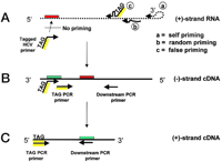
Early attempts to detect negative-strand RNA in vivo and in vitro used a primer specific for the negative strand during the initial reverse-transcriptase step. The reverse transcriptase was then inactivated, and the synthesized DNA was amplified by PCR. Using this technique, negative-strand RNA was found in many tissues and in vitro infected cells. However, this technique was shown to lack strand specificity because of a combination of factors, the most important of which included false priming of the incorrect strand, self-priming of the RNA, and random priming of the RNA by extraneous nucleic acids. This false priming was significant because the reverse-transcription reactions were generally performed at temperatures below 42°C (Fig. 1). These problems were recognized by a number of scientists,12,13 and more reliable techniques were introduced. False priming by extraneous nucleic acids could be avoided by chemically blocking the free 3′ ends of the RNA with borohydride.14 Under these conditions, only the added primer was capable of being elongated by the polymerase. Lanford et al.15 introduced a method termed “tagged” RT-PCR in which the primer used during cDNA synthesis contained a non-HCV (“tag”) sequence at the 5′ end. After reverse transcription, PCR is performed by using a primer corresponding to the “tag” sequence and an HCV-specific oligonucleotide as the opposing primer. When properly used, this technique can yield a 10,000-fold discrimination between detection of plus- and minus-strand RNAs. However, although a huge advance, this technique is still not without problems. Lerat et al.16 showed that specificity was lower than with conventional primers for the HCV core region, although specificity was increased with primers representing the 5′ nontranslated segment of the HCV genome. Nonetheless, recently it was reported that the use of tagged core primers led to a highly strand-specific assay.17
|
|
Fig. 1. Potential causes of nonspecific amplification of positive-strand RNA sequence by using tagged RT-PCR. (A) Conventional reverse transcription of RNA into cDNA at 42°C with the minus-strand specific tagged HCV primer can lead to nonspecific synthesis of cDNA from positive-strand RNA by virtue of (a) self-priming, (b) random prining, or (c) false priming. (B) These nonspecific cDNAs along with some of the tagged primer will be transferred into the subsequent PCR reaction mix. The tagged HCV primer (through its HCV-complementary sequence) can now use the nonspecifically amplified cDNA as template during the first cycle of the PCR reaction. (C) In further PCR cycles, amplification of the target by the PCR primers (tag primer, which lacks HCV sequence, and the downstream HCV primer) leads to a false-positive result. RNA is shown as dotted lines, and DNA as solid lines. “Active” primer sites appear green, whereas “inactive” primer sites are red. The tag sequence is shown in yellow. |
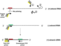
A limitation of this method, perhaps not widely appreciated, is the requirement for the cDNA primer to be exhausted during the reverse-transcription step and for the reverse transcriptase to be fully inactivated. The latter is usually accomplished by heating the reaction at 100°C for 1 hour before the PCR step. An alternative technique, introduced in the same report as the tagged method,15 and which is becoming increasingly popular, is the rTth RT-PCR assay. Here, false priming is prevented by using rTth reverse transcriptase at 70°C. The reverse-transcriptase activity of the enzyme is then inactivated by chelation of Mn2+, and the DNA-dependent polymerase activity of the enzyme is activated by addition of Mg2+ (Fig. 2). Analysis of synthetic RNAs routinely shows at least a 10,000-fold discrimination between the detection of the correct and incorrect strands of HCV in this type of assay.15,18
|
|
Fig. 2. Improved specificity of rTth RP-PCR for detection of minus-strand RNA. (A) Reverse transcription of RNA into cDNA is performed at 70°C in the presence of Mn2+. Because of the higher temperature, there is greatly reduced (a) self-priming, (b) random priming, or (c) false priming. Thus, there is very little nonspecific cDNA synthesized and transferred into the PCR reaction. This results in 1,000- to 10,000-fold specificity for detection of the minus-strand RNA. (B) Correct detection of minus-strand RNA. As in (A), reverse transcription of RNA into cDNA is performed at 70°C in the presence of Mn2+. The tagged HCV primer results in synthesis of a positive-strand cDNA. In the subsequent PCR reaction, the reverse-transcriptase activity of the rTth polymerase is eliminated by the addition of ethyleneglycoltetraacetic acid and Mg2+ is added to activate the DNA polymerase activity. The positive-strand cDNA is now amplified by the tag primer (through its tag sequence) and the downstream HCV primer. |
The identification of HCV as an important pathogen and the importance of being able to reliably detect minute amounts of either strand of RNA have led to significant improvements in RT-PCR technology. Reliable results can be produced with available technologies, provided that a few precautions are performed. Nested RT-PCR should be a “last resort.” Sufficient sensitivity is possible using single-cycle RT-PCR. For specific detection of minus-strand RNA, we recommend using rTth reverse transcriptase; the use of a tagged primer, after determining the optimum level of cDNA primer, will probably lead to a further increase in specificity but at the expense of sensitivity. The optimal target area for specificity must be identified, and the system must be evaluated for specificity by using a full range (over at least 6 logs) of synthetic plus- and minus-strand RNAs, preferably in the environment of total cellular RNA. Blocking the 3′ end of all but the specific primer should be considered as a further precaution.14 A detailed description of the rTth method has recently been published.19
By using some of the above suggestions, many earlier conflicting results are being resolved. It was established in early work that the liver was the primary site for the replication of HCV,20,21 a finding that has been confirmed in many subsequent studies. However, the identification of extrahepatic sites of HCV replication has been more controversial. There have been many reports of the detection of replicative intermediates in peripheral blood mononuclear cells (PBMCs).22 A recent publication reported that 86% of PBMCs that contain positive-strand RNA also contained negative-strand RNA, using in situ RT-PCR.23 However, Mellor et al.17 could find negative-strand RNA in only 1 of 10 dendritic cell preparations taken from chronically infected patients. In the other 9 patients, negative-strand RNA could not be detected in any PBMC fraction. Other workers have also failed to detect Hepatitis C in PBMCs.24,25 Therefore, it appears that if PBMCs are reservoirs of infection, these cells constitute a very small number of infected cells compared with the liver.
Hepatitis C RNA has also been detected in a wide range of other tissues.26,27The strand specificity question has been well addressed in one of these studies. However, confirmation of this work is required before the significance of these results can be judged. The situation in vitro is also controversial. It is well established that primary chimpanzee hepatocytes can be infected,15 and there is a large body of evidence that various lymphoblastoid cell lines are susceptible to HCV infection.28,29 The length of time that these lines continue to produce both positive and negative strands, plus the finding that the quasispecies present within the inoculum are selected to a more limited spectrum after in vitro passage, indicate that replication is indeed taking place. However, it occurs at only a very low level and probably involves only a very small fraction of the cells. It is hoped that by adopting the precautions highlighted above, these studies will be reproduced in other laboratories, which will hopefully point the way towards a truly permissive system for the in vitro culture of HCV. One important aspect of RT-PCR not considered in this article is that of accurate quantitation. With improved technology this goal becomes more realistic, and perhaps the time has come for claims of HCV replication to be supported by accurate quantitative data.
The situation with GBV-C/HGV is even more confusing. The levels of genome equivalents detected in plasma are high, and titers of 107/mL are not uncommon. However, most reports show only low levels of virus in liver samples,17,18 with no evidence of negative-strand RNA. These results are consistent with the finding that GBV-C/HGV infection does not correlate with overt liver disease in patients on maintenance hemodialysis.30 Similarly, attempts to detect virus in PBMCs have generally been negative. Thus, although it is clear that GBV-C/HGV is replicating to high levels in some tissue, it is by no means clear what this tissue is. Perhaps the name “Hepatitis G virus” should be dropped completely in favor of the alternative GBV-C or “human orphan flavivirus” as suggested by Theodore and Lemon.31
In summary, RT-PCR has been invaluable in the investigation of Hepatitis C and the more recently identified GB viruses. However, its extreme sensitivity has led to problems both with respect to false positives and the differentiation of positive- and negative-strand RNAs. These problems are now well recognized. With attention to a small number of important precautions, it should be possible to move forward in further elucidating the role of these viruses to human disease.
Acknowledgment
The authors are grateful to Dr. Stanley Lemon for advice, for carefully reading the script, and for making numerous suggestions.
Abbreviations
Abbreviations: PCR, polymerase chain reaction; RT-PCR, reverse-transcriptase polymerase chain reaction; cDNA, complementary DNA; HCV, Hepatitis C virus; PBMC, peripheral blood mononuclear cell.
Footnotes
Received June 15, 1998; accepted August 14, 1998.
Address reprint requests to: David V. Sangar, Ph.D., Department of Microbiology and Immunology, University of Texas Medical Branch at Galveston, Galveston, TX 77555-1019. Fax: (409) 772-3757; e-mail: dvsangar@utmb.edu.


